I am currently preparing to shoot in the lab at the end of the month, and am also in the midst of a large data/image collection, part of developing two works for a public installation. The image experimentation is a case of aligning and connecting workflows from scientific imaging and media, and testing and re-testing the samples and their imaging methods and aesthetics.
Here are a couple of examples of the compiled videos I have been working with – they’re not at their full resolution just yet — I plan to scale these up over the coming weeks to increase post-production flexibility. More to come soon.
Lung Tumour cells. These are used in the lab to explore the use of nanoparticles in imaging systems that might help to detect tumour cells in the human body.
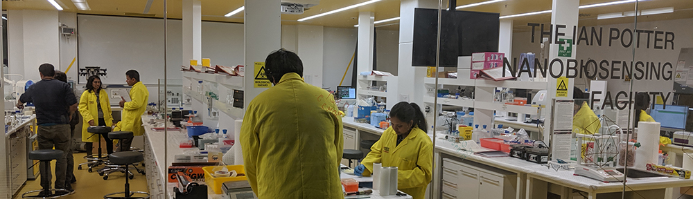
The Ian Potter NanoBioSensing Facility is developing light-activated sensing systems for applications such as self-cleaning clothes and glucose or gas sensors. They investigate the light-activation of nanoscale compounds bound onto fabrics. The chemists deposit copper, platinum, palladium or gold nanoparticles onto cotton fabric and then analyse the way light activates the electron transfer process between the nanoparticles and the molecules they are detecting. Light is absorbed by the nanoparticles in the presence of a target molecule, for example glucose. The system then undergoes a colour change, giving a simple visual signal of the presence of glucose. The example above is cotton fabric coated with copper and then palladium nanoparticles (not visible here).